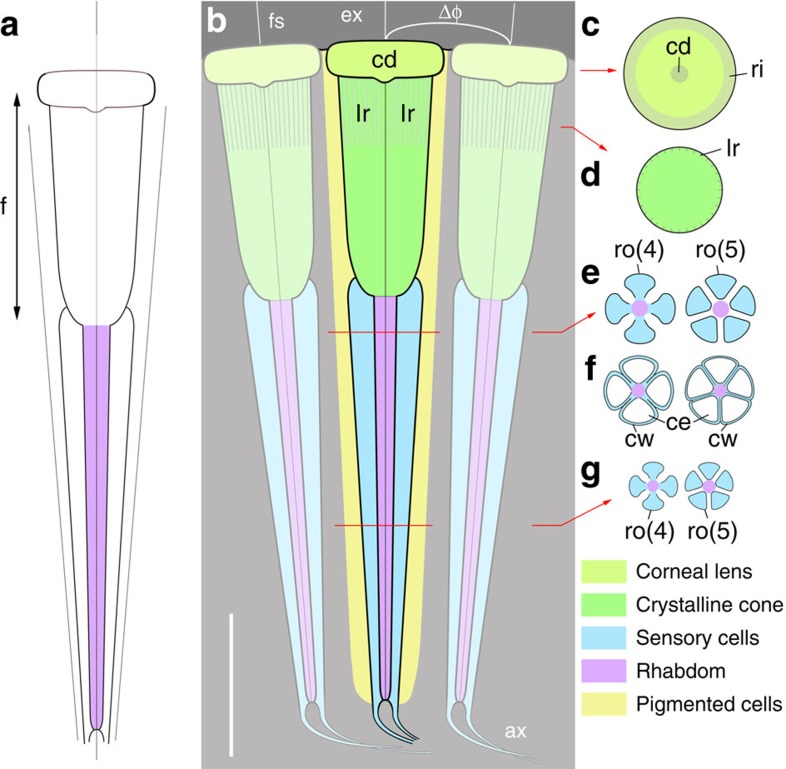
Figure 3. Reconstruction of the eye structure of Dollocaris.
Thylacocephala; Middle Jurassic, showing three adjacent ommatidia. The most distal disk-like feature is interpreted as the corneal lens. The underlying crystalline cone is assumed to form an image at its proximal tip in direct contact with the rhabdom. The 4 or 5 retinula cells form a long tapering cylindrical feature that extends into nerve fibres. In transverse sections, the retinula cell clusters appear as a rosette-like structure with the central axis probably representing the rhabdom. Cellular details of the inter-ommatidial region are not preserved except possible pigmented areas along the crystalline cone (Supplementary Fig. 3). (a) Simplified ommatidium. (b–g) Simplified longitudinal (b) and transverse sections through ommatidia with 4 and 5 retinula cells and two types of cellular preservation (cells empty with mineralized cellular walls or cells mineralized as a whole). ax, axonal structure; cd, central depression; ce, cell; cw, cell wall; d, rhabdom diameter; Δφ, inter-ommatidial angle; ex, external medium; f, focal length; fs, faceted surface; lr, longitudinal ridge; ri, rim; ro(4), rosette-like structure with 4 cells; ro(5), rosette-like structure with 5 cells. Scale bar, 50 μm.

