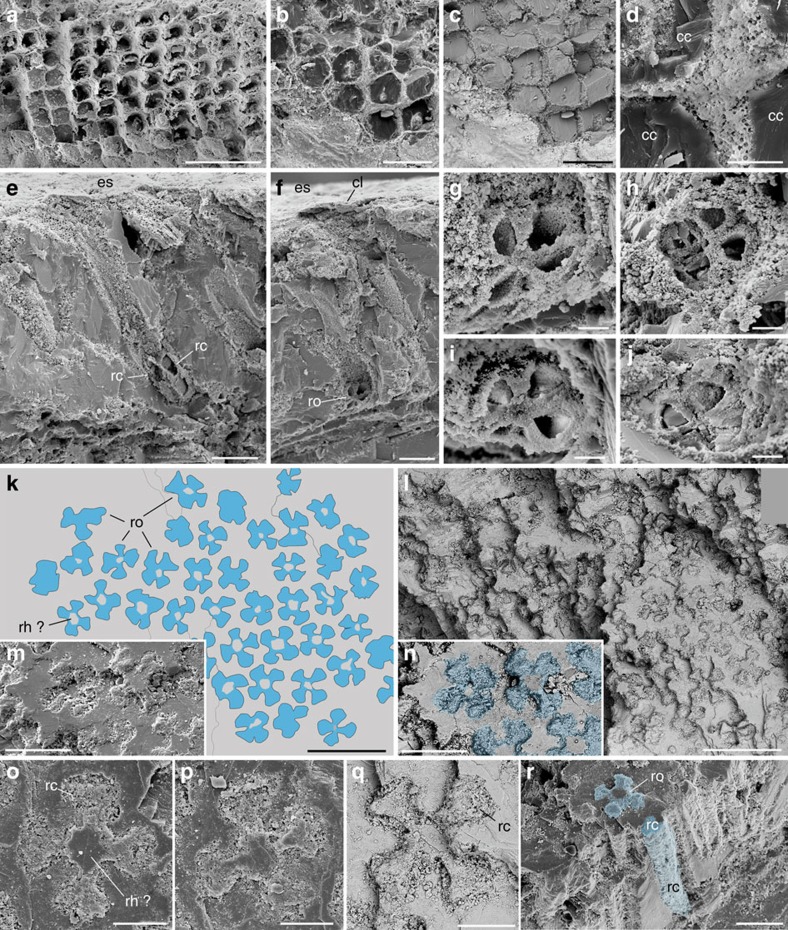
Figure 4. Internal structure of the eye of Dollocaris ingens.
Thylacocephala; Middle Jurassic La Voulte Lagerstätte. (a–d) Transverse sections through adjacent ommatidia showing juxtaposed crystalline cones, general view, details and interspace between crystalline cones. (e) Longitudinal section through ommatidia showing two retinula cells. (f,g) Transverse section through a rosette-like cluster of 4, possibly 5 retinula cells, general view and details. (h–j) Details of rosette-like clusters with 4 retinula cells. (k–n) Transverse section through the retinula cells of numerous ommatidia, general view, simplified drawing (cells in blue) and details. (o–q) Details of rosette-like structures. (r) Rosette-like structure and elongated three-dimensional structure of a retinula cell. FSL 710064 in a–j, MNHN.F.R06206 in k–r. All SEM images. c,l,n,q are back-scattered images. cc, crystalline cone; cl, corneal lens; es, eye external surface; rc, retinula cell; rh?, possible rhabdom; ro, rosette-like structure (section through cluster of retinula cells). Scale bars, 100 μm in a,k,l; 50 μm in b–d,m,n,r, 20 μm in e,f,o–q, 5 μm in g–j.

