Figure 4.
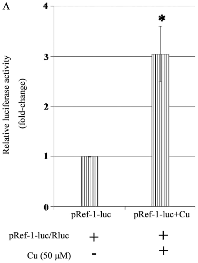
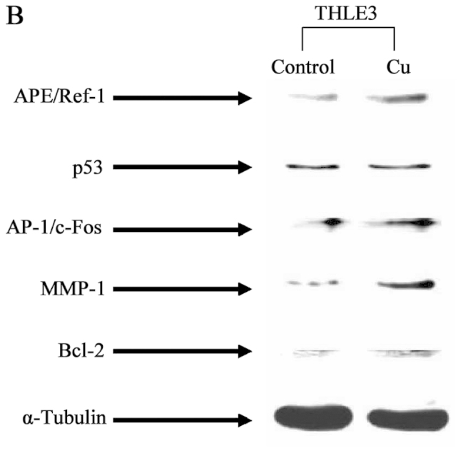
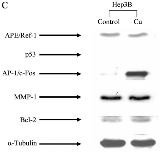
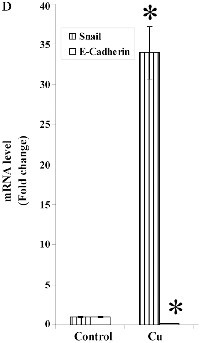
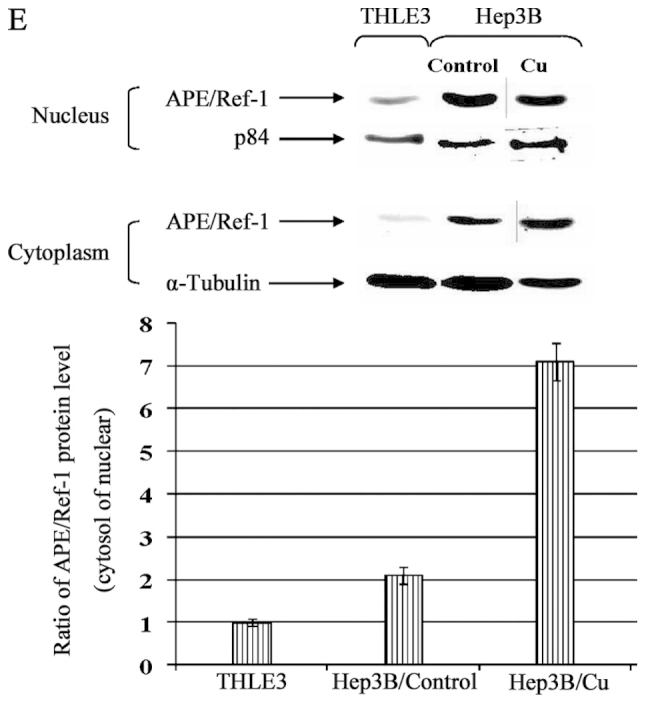
Cu treatment induced APE/Ref-1 as well as its target genes in THLE3 hepatocytes and Hep3B cells. (A) Cu potentiates promoter activity of APE/Ref-1 which was determined by Dual Luciferase Reporter assay. Induction of APE/Ref-1, AP-1/c-Fos, MMP-1 and Bcl-2 by Cu treatment in THLE3 (B) and Hep3B cells (C). (D) Quantitative real-time RT-PCR analysis of Snail and E-cadherin in Hep3B cells following 24-h incubation with Cu 50 μM. (E) Alteration of APE/Ref-1 sub-localization quantified by the ratio of cytoplasmic/nuclear APE/Ref-1 relative optical density values. Following 24-h incubation with Cu 50 μM, cells were collected and lysed. Equal amounts of the soluble protein were subject to western blotting. For statistical analysis, all data were expressed as fold changes of the control based on the calculation as the density values of the specific protein band/α-tubulin or p84 density values. Data are expressed as fold change. *p<0.05; **p<0.01 versus control. Columns, mean (n=3); bars, SD.
