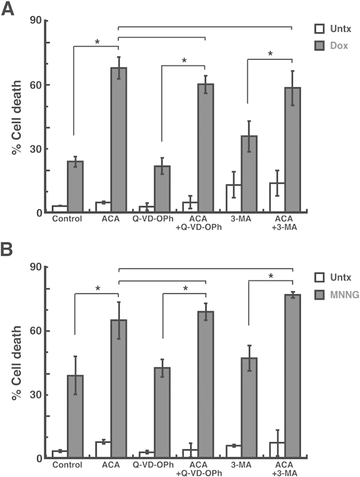
Figure 4.
Analysis of apoptosis and autophagy in breast adenocarcinoma cells after TRPM2 inhibition and chemotherapeutic treatments. (A) MDA-MB-231 breast adenocarcinoma cells were pretreated with 20 μM ACA for 30 min, and then treated with 2 μM doxorubicin (Dox). Cell death was then quantified by flow cytometry. For analysis of apoptosis, cells were pretreated with 50 μM Q-VD-OPh, a pan-caspase inhibitor. For analysis of autophagy, cells were pretreated with 3-methyladenine (3-MA), an inhibitor of autophagy. (B) MDA-MB-231 breast adenocarcinoma cells were treated as in A, except a 30-min exposure to 100 μM MNNG was used as the chemotherapeutic treatment. *p<0.05, one-way ANOVA and unpaired Student's t-test; error bars represent the SEM.

