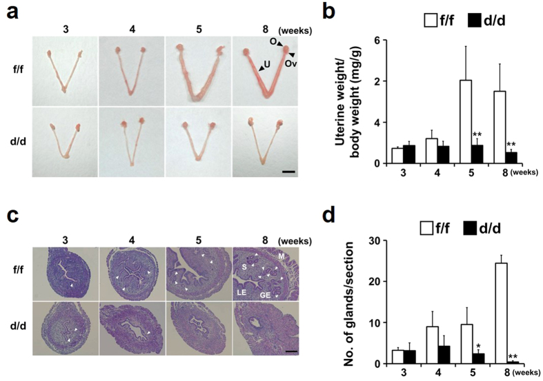
Figure 4. Gross and histological analyses for uterine development of Dgcr8d/d mice at various ages.
(a) Gross morphology of the uteri of 3- to 8-week-old Dgcr8d/d mice. Note that while the uteri of 3-week-old Dgcr8d/d mice look normal, they become severely abnormal beginning at 5 weeks of age. Scale bar: 5 mm. (b) Changes of uterine weight/total body weight of Dgcr8d/d mice during uterine development. Consistent with the distinct morphologic abnormalities observed in mice beginning at 5 weeks of age, uterine weight was dramatically lower than that of control from 5-week-old stage (n = 4 to 6 for each group). (c) Representative microscopic images of the uteri of 3- to 8-week-old Dgcr8f/f and Dgcr8d/d mice. Scale bar: 100 μm. (d) The number of uterine glands/section observed in Dgcr8f/f and Dgcr8d/d mice (n = 4 to 6 for each group). Unpaired Student’s t-test, *p < 0.05, **p < 0.01. O, Ovary; Ov, Oviduct; U, Uterus; M, Myometrium; S, Stroma; GE, Gland epithelium; LE, Luminal epithelium. White arrowheads indicate glands.

