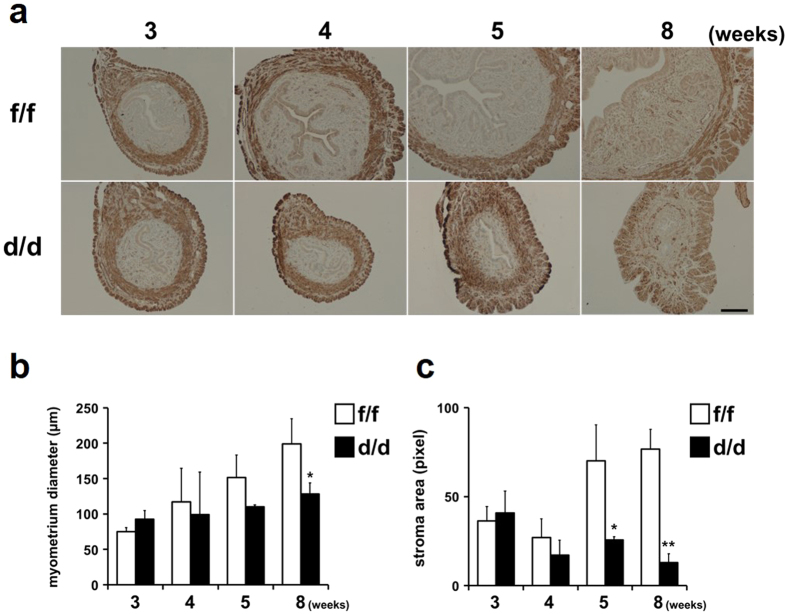
Figure 5. Stromal and myometrial abnormalities during uterine development of Dgcr8d/d mice.
(a) Immunohistochemical analyses for alpha smooth muscle actin (α-SMA) in the uterine myometrial layers of Dgcr8d/d mice. Dark brown color indicates α-SMA positive smooth muscle cells in the uterus. Scale bar: 100 μm. (b) Myometrial thickness in the uteri of Dgcr8f/f and Dgcr8d/d mice (n = 4 to 6 for each group). Myometrial thickness was determined by the length of the area of α-SMA positive cell layers. (c) Quantification of stromal area during uterine development in Dgcr8d/d mice. Stromal area was quantitatively measured by pixels of images for each uterine section of Dgcr8f/f and Dgcr8d/d mice. At least three independent sections were microscopically examined for each mouse (n = 4 to 6 mice for each group). Unpaired Student’s t-test, *p < 0.05, **p < 0.01.

