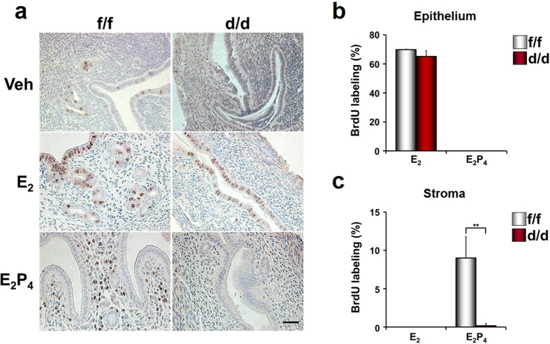
Figure 6. Cell proliferation in the uteri of ovariectomized Dgcr8d/d mice treated with ovarian steroid hormones.
(a) BrdU incorporation experiments were performed to examine hormone-dependent endometrial cell proliferation in Dgcr8d/d mice. Ovariectomized mice were sacrificed 24 h after injection with vehicle, E2 or E2 + P4. BrdU was given to these mice 3 h before sacrifice. Brown color indicates the nuclei of BrdU-incorporated cells. Scale bar: 25 μm. (b,c) Graphs depicting the percentage of BrdU positive cells/total number of cells counted. Note that stromal cell proliferation was severely impaired in Dgcr8d/d mice, whereas uterine epithelial cells normally responded to E2. Unpaired Student’s t-test, *p < 0.05, **p < 0.01.

