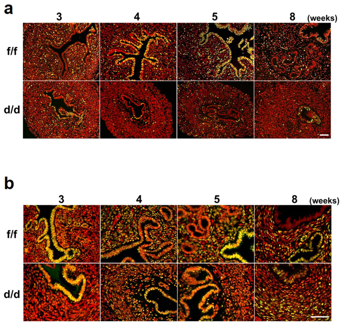
Figure 7. Expression of Ki-67 and PR in uteri of Dgcr8d/d mice at various stages.
(a,b) Immunofluorescence of Ki-67 (a) and PR (b) on uterine sections from Dgcr8f/f and Dgcr8d/d mice at various ages. Ki-67 or PR was visualized as green and nuclei were stained with TO-PRO-3-Iodide (red). Yellow color indicates Ki-67 or PR positive cells. Scale bar: 50 μm.

