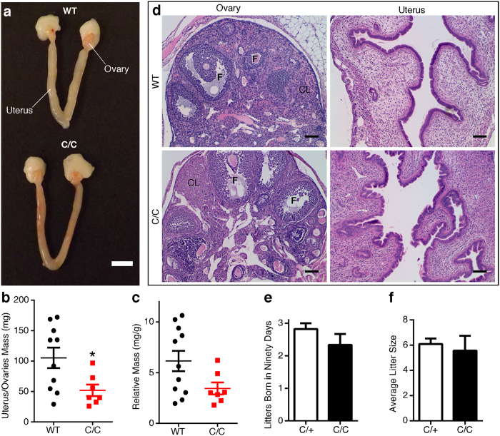
Figure 2. P42 female C/C reproductive organs exhibit minimal abnormalities.
(a) Representative photograph of WT (top) and C/C (bottom) uterus and ovaries. Scale bar represents 5 mm. (b) Gross uterus and ovaries mass. (c) Relative uterus and ovaries mass was determined by dividing gross uterus and ovaries mass by total body weight (n = 10 WT and 7 C/C mice). (d) Representative H&E stained cross-sections of ovary and the endometrium. Follicle (F) and corpus luteum (CL) are indicated on ovary micrographs. Scale bars represent 100 μm. (e–f) Fertility of heterozygous (n = 11) and C/C (n = 3) female mice. Females were paired in monogamous breeding cages with either a heterozygote or WT male. The average number of litters born over ninety days (e) and the average litter size for heterozygote or C/C mothers (f) were recorded. (Statistical significance *p < 0.05).

