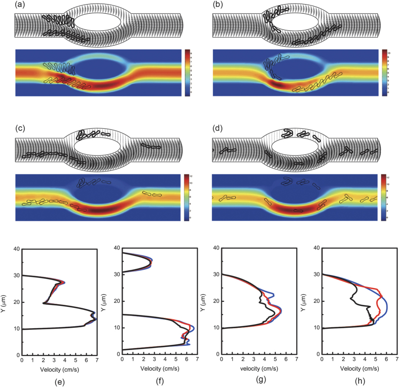
Figure 7.
Motion of two files of 44 red blood cells (Hct = 17.6%) in the asymmetric bifurcated microchannel at time instants (a) t = 1.20 ms, (b) t = 1.80 ms, (c) t = 2.40 ms, and (d) t = 5.00 ms. Velocity vectors (upper panels) and axial velocity magnitude contours (cm/s) (lower panels) are presented. The reduced area s* = 0.481. The spring constant of the cell membrane was kl = kb = 3.0 × 10⁻¹³ Nm. Velocity profiles at different locations of the microvessel for different hematocrits: (e) at the apex of the diverging bifurcation at t = 1.20 ms, (f) at the mid cross section of the bifurcation at t = 1.80 ms, (g) at the apex of the converging bifurcation at t = 2.40 ms, and (h) at the cross section 2 μm from the right outlet at t = 5.00 ms. Blue line: 8 red blood cells (Hct = 3.2%); red line: 16 red blood cells (Hct = 6.4%); black line: 44 red blood cells (Hct = 17.6%); green line: 80 red blood cells (Hct = 32%).

