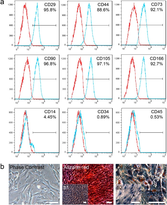
Figure 1. Characterization of UC-MSCs.
(a) Immunophenotype of isolated UC-MSCs. Isolated UC-MSCs were characterized by FACS. UC-MSCs were positive for CD29, CD44, CD73, CD90, CD105, and CD166, and nearly negative for CD14, CD34, and CD45. (b) Differentiation characteristics of UC-MSCs. The phase contrast of UC-MSCs is shown on the left. Osteogenic differentiation was detected by Alizarin red staining (middle), and adipogenic differentiation was visualized by Oil Red O staining of the lipid vesicles (right). UC-MSCs cultured with normal medium were used as the negative control group for each stain (b1 and b2). All scale bars correspond to 100 μm.

