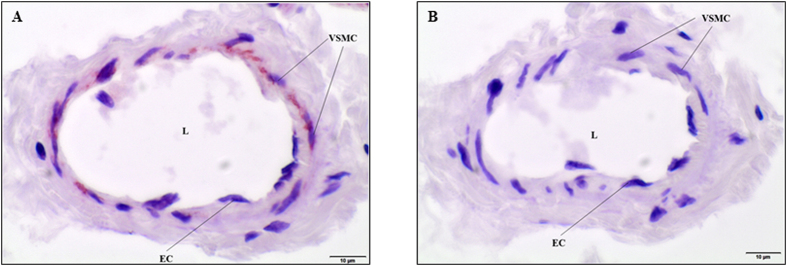
Figure 7. Photomicrographs representing the transverse cryosections of the mouse ophthalmic artery for Kv1.6 channel immunolocalization.
(A) The expression of the Kv1.6 channel is prominent in the smooth muscle cell layer compared to the endothelial cells. (B) The negative control demonstrates no staining in the absence of the primary antibody. EC, Endothelial cell; VSMC, Vascular smooth muscle cell; L, Lumen. Scale bars indicate 10 μm at 600× magnification.

