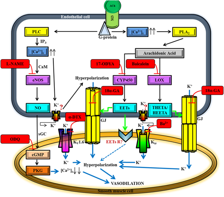
Figure 9. The proposed hypothetical endothelial cell-dependent signalling pathways of conducted vasodilation in response to ACh in the mouse ophthalmic artery.
The first proposed mechanism involves the NO/cGMP pathway: Activation of the endothelial M3 receptor by ACh induces an influx of [Ca2+]i. Following interaction with CaM (calmodulin), Ca2+ activates eNOS and release of NO. NO causes relaxation by interacting with the haem group of the enzyme, sGC, which then mediates the formation of cyclic guanosine monophosphate (cGMP) and activation of protein kinase G (PKG) that relaxes the VSMC. The second vasodilator mechanism is via the arachidonic acid (AA) metabolites synthesized through the CYP450 oxygenase pathway: The increase in [Ca2+]i elicits translocation of phospholipase A2 (PLA2) to the membrane and its major hydrolysis product is AA, which can be metabolized by CYP450 oxygenase to EETs. The EETs function as messenger molecules that modulate gap junctions (GJ) to spread the conductance to the VSMC. EETs may also directly activate a channel/receptor on the VSMC to hyperpolarize and dilate the vessel. The third signalling pathway involves the AA metabolites generated via the LOX pathway: It is hypothesized that LOX and/or its metabolites, namely THETA and HEETA do not pass the GJ as EDHF per se but activate GJ to hyperpolarize the VSMC. The forth key players are the gap junctions. The fifth proposed mechanism involves the active participation of the Kir and Kv1.6 channels: It is hypothesized that both Kir and Kv1.6 channels on the VSMC are activated by the increase in extracellular K+ resulting in hyperpolarization and vasodilation. The precise identity of the putative channel(s) on the endothelial cells that is activated and opened for K+ efflux for hyperpolarization to occur is unknown. Question marks represent unknown receptors that are yet to be identified. Blockers and inhibitors are indicated in red boxes. Green arrows indicate the activation of the gap junctions and downstream receptor(s). Blue solid arrows show potential pathways for transfer of hyperpolarization from the endothelium to the smooth muscle cells. Blue quadrangular point arrow indicates the hypothesized transfer of hyperpolarization via an unknown receptor on the VSMC.

