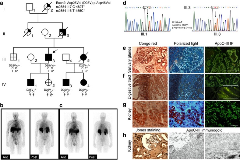
Figure 1. Apolipoprotein C-III amyloidosis in a French family.
(a) Pedigree showing three generations of transmission of the ApoC3 D25V variant with systemic amyloidosis and decreased plasma triglyceride levels:  APOC3 mutation present;
  clinical syndrome;
  ApoC-III amyloid confirmed histologically;
  decreased plasma triglyceride levels;
  no clinical syndrome, no amyloid on histology, absence of APOC3 mutation, no hypotriglyceridemia. APOC3 haplotypes distribution of C-482T and T-455C polymorphisms is shown. D25V+/−, heterozygous for the apoC-III amyloidogenic variant; D25V−/−, absence of apoC-III mutation. (b,c) Serial anterior and posterior planar whole-body 123I- SAP scan. The scans from III.3 in b and IV.3 in c show a congested liver and amyloid in the spleen and kidneys. (d) Partial sequence chromatograms of APOC3 exon 3. (e) Amyloid deposits in salivary glands from proband III.3. (f) Amyloid deposits in the duodenum from proband III.3 predominate in the submucosa around the vessels and around Brenner's glands. There are also amyloid deposits grouped in small clods within the intestinal villi. (g) Abundant vascular, moderate glomerular and interstitial amyloid deposits in kidney from proband III.3. For each tissue in e–g, left panels show the Congo red staining, scale bar, 100 μm; middle panels show the Congo red-stained section viewed under polarized light, scale bar, 100 μm; right panels show the immunofluorescence with anti-apoC-III antibody; scale bar, 100 μm. (h) Left panel: amyloid deposits in the kidney are observed in the mesangium and the glomerular arteriole (arrow) (Jones staining, scale bar, 100 μm); middle and right panels: electron micrograph of amyloid fibrils on immunogold staining with anti-apoC-III antibody, scale bar, 500 and 200 nm, respectively).

