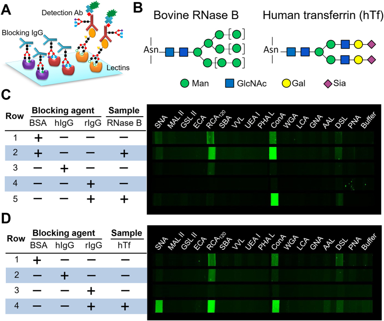
Figure 3. Development and characterization of the microfluidic antibody-lectin sandwich barcode assay.
(A) Binding of the glycosylated detection antibody on the arrayed lectins can cause severe non-specific background. To mitigate this effect, the target-bound lectin array is blocked with a non-labeled IgG blocker prior to detection using a biotinylated antibody and fluorescently labeled streptavidin. (B) The predominant glycan structures on the standard glycoproteins used: bovine RNase B and human transferrin (hTf). The square brackets indicate the possible high mannose variants on RNase B. (C,D) False-color fluorescence images of glyco-profiling of bovine RNase B (C) and hTf (D) using a 16-lectin barcode array and different blockers-BSA, human plasma IgG (hIgG) and rabbit serum IgG (rIgG). The concentrations of RNase B and hTf are 5 and 50 μg/mL, respectively.

