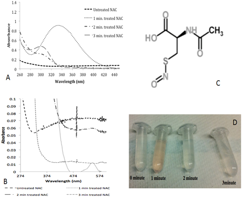
Figure 2. UV-Visible Spectra of Plasma Treated NAC Solution.
(A) Following 1-minute plasma treatment of NAC solution, a peak at 332 nm (representing formation of S-nitroso N-acetyl cysteine (S-NAC); a type of S-nitrosothiol molecule) was observed. Following 2-minute of plasma treatment, the peak at 332 nm was shifted to 302 nm and the intensity of the peak at 302 nm increased in the plasma treatment time dependent manner. (B) Specific secondary peak for S-nitroso N-acetyl cysteine (S-NAC) in 1-minute plasma treated NAC solution. (C) S-nitroso N-acetyl cysteine (S-NAC) molecule, H atom is abstracted from thiol group and NO group is bonded after 1-minute of plasma treatment. (D) A change in color was observed in 1-minute plasma treated NAC solution, which is characteristic for s-nitrosothiols. Our observation on plasma treated NAC solution along with UV-vis results suggests the formation of S-NAC in 1-minute plasma treated NAC solution.

