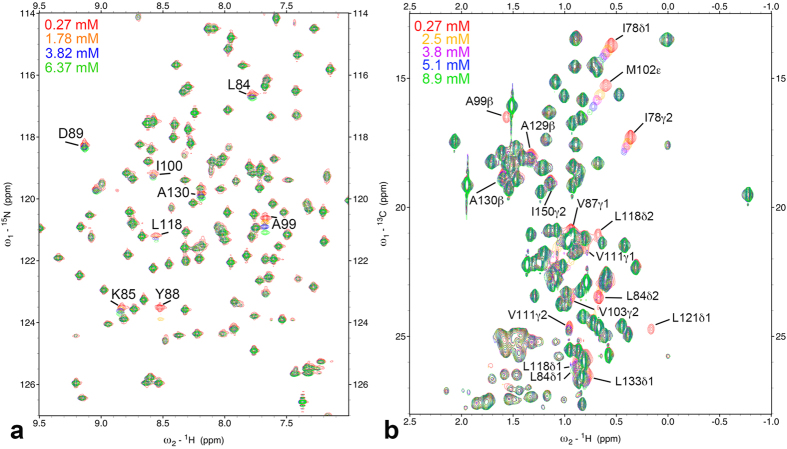
Figure 1.
(a) 1H/15N refocused-HSQC spectra of 15N labeled L99A of T4 lysozyme at 298 K at different oxygen concentrations from 0.27 mM to 6.4 mM. Amide groups showing significant changes in 15N chemical shift are indicated. (b) 1H/13C constant time HSQC spectra of 13C/15N labeled L99A of T4 lysozyme at different oxygen concentrations from 0.27 mM to 8.9 mM. Positive and negative crosspeaks are presented by same color. Methyl groups showing significant changes in 1H/13C chemical shift and a loss of crosspeak intensities are indicated.

