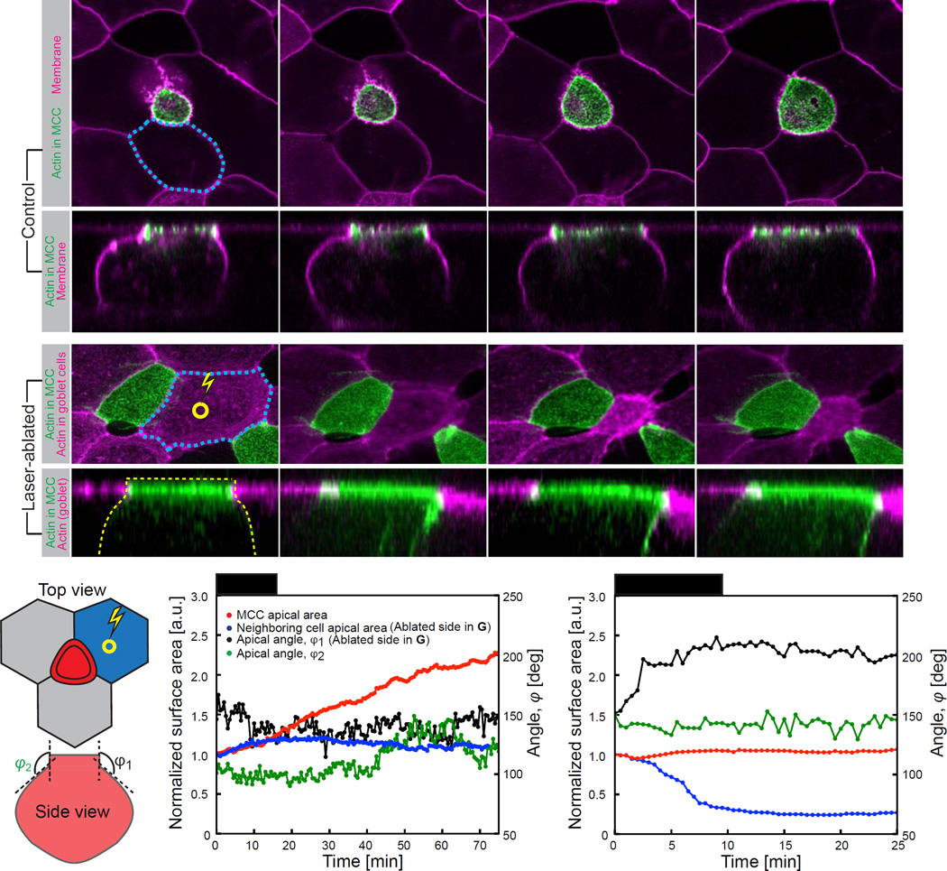
Figure 2. Puling forces of cells adjacent to MCCs do not drive apical emergence process.
(A) Image sequence of apically expanding MCC expressing actin marker – utrophin (UtrCH, green) under MCC specific α-tubulin promoter; epithelial cells visualized by expression of membrane marker CAAX-RFP mRNA (magenta). (B) Orthogonal projections, corresponding to (A). (C) Image sequence of MCC apical domain upon laser ablation in the center (indicated by yellow circle) of the neighboring goblet cell. Laser ablation leads to excessive constriction of goblet cell (outlined in blue dotted line) but does not result in MCC apical domain collapse. (D) Orthogonal projections, corresponding to (C). Constricting goblet cell exerts pulling force on the MCC apical domain, as assessed by change of the angle between apical and lateral side of MCC. (E) Parameters measured upon laser ablation of goblet cell adjacent to MCC: apical area of ablated goblet cell, blue; apical area of MCC adjacent to ablated goblet cell, red; angles between apical and lateral side, φ1 (ablated side) and φ2 (non-ablated-side). (F) Dynamics of parameters described in (E) in control and upon laser ablation in the center of the goblet cell (G). Despite the evident pulling by the neighboring goblet cell (increase of φ1 in (G)), apical area of MCC does not increase, compare to (F). Scale bar, 10µm., a.u., arbitrary units. See also Figure S3.

