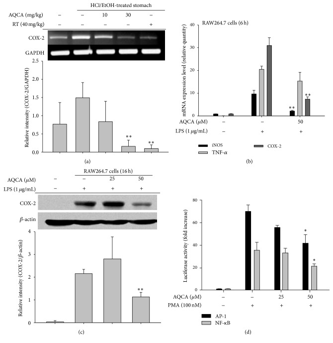
Figure 4.
Effect of AQCA on the transcriptional activation of inflammatory genes. (a and b) mRNA levels of inflammatory genes (iNOS, TNF-α, and COX-2) in mice treated with HCl/EtOH or LPS-treated RAW264.7 cells were determined by semiquantitative (a) and real-time RT-PCR (b). (c) Total lysates were prepared from LPS-treated RAW264.7 cells treated with AQCA (25 and 50 μM). The total forms of COX-2 and β-actin were analyzed by immunoblotting analysis. (d) Promoter binding activities of NF-κB and AP-1 in the presence or absence of PMA were determined by reporter gene (luciferase) assay. Relative intensity (a and c) was calculated using level of GAPDH or β-actin and the DNR Bio-Imaging System (a and c bottom panels). ∗ P < 0.05 and ∗∗ P < 0.01 compared with the control group.

