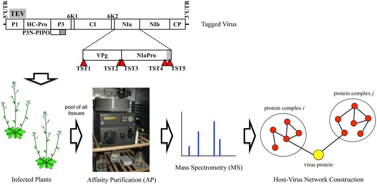
Fig. 1.

Schematic representation of the TEV recombinant clones in which the NIa protein was tagged with the Twin-Strep-tag (TST) at five different positions (TEV-TSTNIa1 to TEV-TSTNIa5) and affinity purification (AP) mass spectrometry (MS) workflow to construct NIa-host interaction networks. In virus scheme, black lines represent TEV 5′ and 3′ UTR, as indicated. Boxes represent TEV cistrons (P1, HC-Pro, P3, P3N-PIPO, 6K1, CI, 6K2, NIa, VPg, NIaPro, NIb and CP) as indicated. Positions where TST was inserted in the VPg and NIaPro domains of NIa are indicated with red triangles. The gray rectangle in NIaPro represents the carboxy-terminal polypeptide that is subjected to processing through a suboptimal autoproteolytic site
