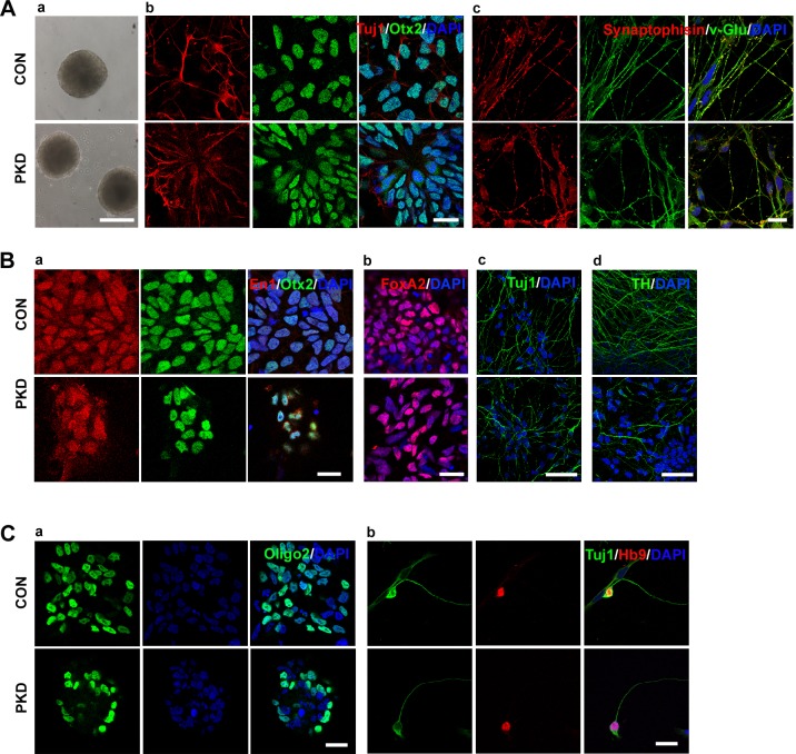
Fig. 4.
Urinary cell-derived iPSCs differentiated into glutamatergic, dopaminergic and motor neurons. (Aa) PKD- and CON-iPSCs formed embryo bodies on the 4th day of differentiation. (Ab) On day 28, induced neurons expressed rostral marker Otx2 (green), and Tuj1 (red). (Ac) Cells were positive for Tuj1 and synaptophysin when differentiated for 7 weeks. (Ba) Cells were positive for midbrain dopamine neuron progenitor markers En1 (red), Otx2 (green) and DAPI (blue) on the 24th day. (Bb) Midbrain dopamine neuron progenitor expressed FoxA2 (red) on the 24th day. (Bc,d) Cells expressed mature neurons marker Tuj1 (Bc) and TH (Bd). (Ca) Immunostaining of motor neuron progenitor marker Olig2. (Cb) Neuronal identity of motor neurons is confirmed by co-expression of HB9 (red) and TuJ1 (green) in dissociated patient-specific motor neuron cultures on the 28th day. Scale bars: 200 µm in Aa; 50 µm in Bc,d; 20 µm in Ab,c, Ba,b and C.

