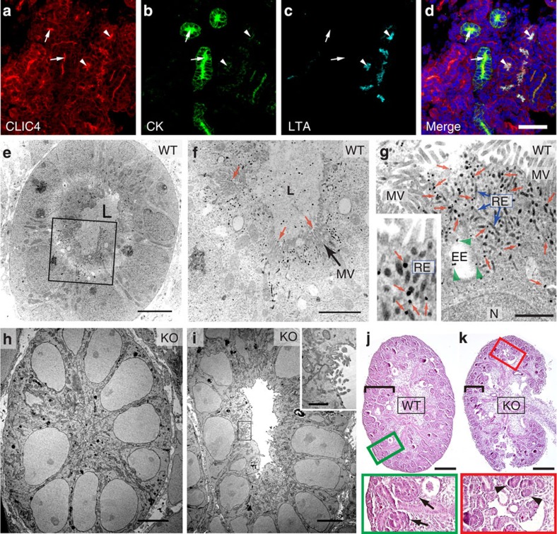
Figure 1. Expression of CLIC4 in embryonic kidney and characterization of CLIC4-KO metanephros.
(a-d) E15 WT metanephros labelled for CLIC4, cytokeratin (CK) and LTA. DAPI: blue. Arrows and arrowheads point to the lumens of cytokeratin and LTA-labelled tubules, respectively. (e-i) Electron micrographs of pre-PT cells of WT (e-g) and CLIC4-KO (h-i) E15 metanephros labelled with CLIC4 immunogolds. The metanephros lumen (L) in WT is filled with electron-dense ECM. (f,g) Enlarged views of the boxed area in (e). CLIC4 immunogolds in MV (red arrows in f), EE (green arrowheads in g) and RE (blue arrows in g). The inset highlights the CLIC4 immunogolds (red arrows) on electron-dense RE tubules. N: nuclei. (h) A mutant PT without detectable central lumen. (i) A hypomorphic mutant PT containing an electron-dense ECM-devoid lumen. The inset shows the enlarged view of the boxed luminal surface demonstrating the presence of residual CLIC4 immunogolds on the short, irregular MV-like processes. (j,k) PAS staining of longitudinally sectioned E15 metanephros of WT (j) and CLIC4-KO (k) mice. Black brackets indicate the cortical areas. Enlarged views of boxed areas highlight the primitive tubules (arrows) and condensed mesenchyme clusters (arrowheads) found in the WT and KO, respectively. Scale bars, 50 μm (d); 5 μm (e,h,i); 2 μm (f); 1 μm (g, inset of i); 200 μm (j,k).

