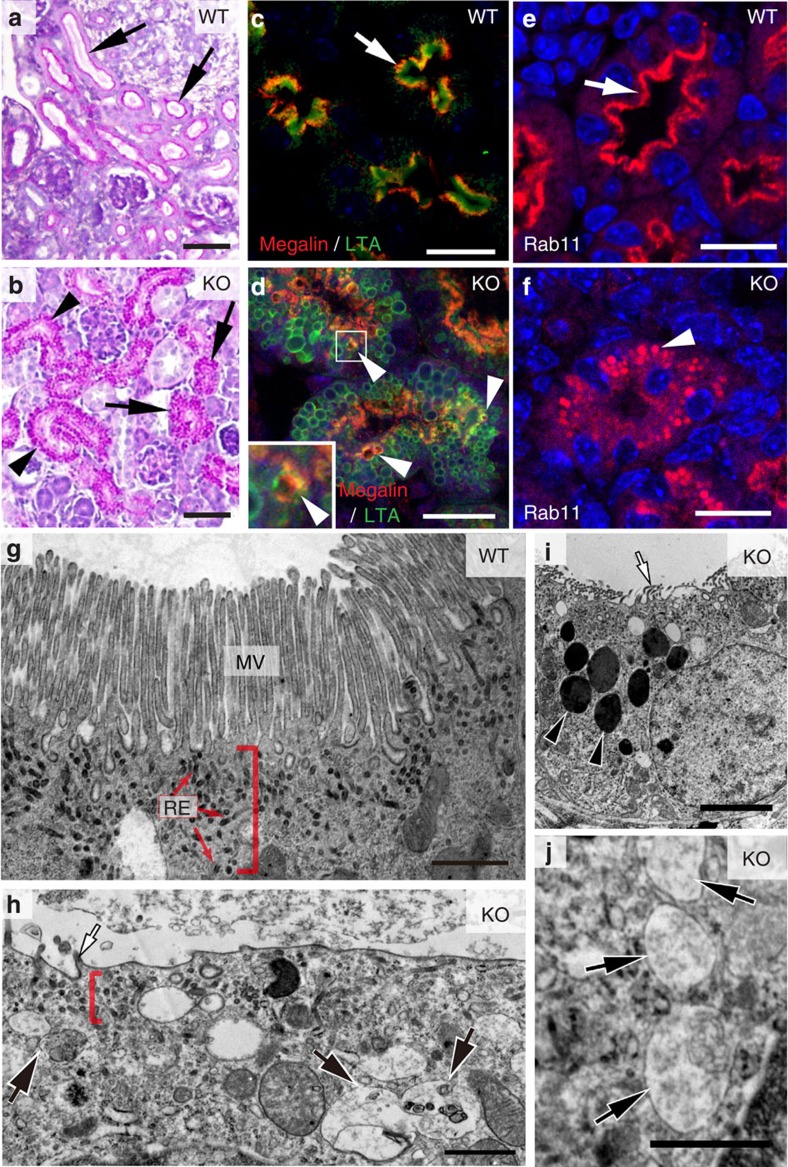
Figure 2. Lumen formation and maturation defect of CLIC4-KO developing PT.
(a,b) PAS-stained renal cortical sections of PN0 WT (a) and CLIC4-KO (b) mice. Arrows in (a) point to the PTs displaying intense PAS+ luminal PM signals. Arrows and arrowheads in (b) point to the cell clusters and the primitive tubes, respectively; both of them contained PAS-positive granules. (c,d) LTA and megalin staining of PTs in PN0 WT(c) and CLIC4-KO (d) mice. Prominent luminal LTA and subluminal megalin signals (arrow) were seen in the WT. Mutant PTs displayed abundant LTA-labelled vacuoles; some contained mislocalized megalin (arrowheads). The inset shows the magnified view of the box area. (e,f) Immunolabelling of Rab11a in PN0 WT (e) and KO (f) PTs. An arrow points to the (sub)luminal lining pattern of Rab11a staining in WT. An arrowhead in (f) points to the granular/vacuolar staining pattern of the remaining Rab11a in mutant PT. (g-j) Representative electron micrographs of PN0 PTs in WT (g) and CLIC4-KO (h-j) mice. Red brackets indicate the apical cytoplasmic regions containing RE (red arrows), which are electron-dense tubules. Mutant PT cells did not develop typical MVs (white arrow, h,i), but contained unusually high numbers of LE-, autophagosome- (black arrows in h,j) and lysosome- (black arrowheads in i) like structures. Scale bars, 50 μm (a,b); 20 μm (c-f); 1 μm (g,h,j); 3 μm (i).

