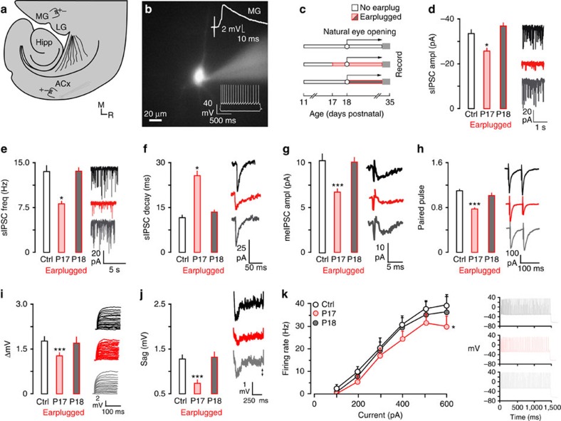
Figure 1. Natural eyelid opening closes the ACx CPs of sensitivity to hearing loss.
(a) Diagram illustrates the thalamocortical brain slice preparation (ACx, auditory cortex; Hipp, hippocampus; LG, lateral geniculate; MG, medial geniculate;). The approximate position of the recording electrode in ACx and the stimulating electrodes in MG and ACx are shown. (b) A fluorescent image of a neurobiotin-labelled pyramidal neuron. An MG-evoked EPSP validated that neurons were located in thalamo-recipient ACx (top right). Representative neuronal responses to injection of current pulses were used to validate a regular pyramidal cell spiking pattern (bottom right). (c) Diagram illustrates the experimental design for data presented in d–k. Earplugs were inserted when eyes were naturally closed or when eyes were naturally opened. Earplugs were maintained until the day of recording (P29 to 35). Sample size; IPSCs: Control, n=40; P17, n=21; P18, n=20; Intrinsic: Control, n=57; P17, n=19; P18, n=24. (d–f) Bar plots (left) and traces (right) show decreased sIPSCs amplitude and frequency, as well as slower decay time constants when ears were plugged before natural eyelid opening (P17), but not after (P18). (g,h) Bar plots (left) and traces (right) show decreased meIPSC amplitude and paired pulse depression when ears were plugged on P17, but not P18. (i–k) Bar plots (left) and traces (right) show that ΔmV, sag and maximum firing rate are decreased when earplugs were inserted on P17, but not P18. Experiments were not replicated. Tukey's (honestly significant different) test, *P<0.05, **P<0.01, ***P<0.001. Error bars are s.e.m.

