Fig. 1.

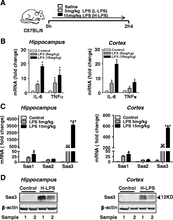
Induced expression of SAA and selected inflammatory cytokines in the mouse brain after systemic administration of LPS. a A schematic representation of experimental design. Three-month-old C57BL/6 mice were injected i.p. with LPS at either 5 mg/kg body weight (L-LPS) or 15 mg/kg body weight (H-LPS) or with same amount of saline (control). Twenty-four hours later, the mice were sacrificed and the hippocampus and cerebral cortex were collected for analysis. b Real-time PCR quantification of the transcripts for IL-6 and TNF-α in the tissue samples of the hippocampus (left) and cortex (right) 24 h after LPS or saline (control) administration. The relative transcript expression was calculated as 2-ΔΔCT and was normalized against saline controls. c relative mRNA levels of Saa1, Saa2, and Saa3 in extracts of hippocampus (left) and cortex (right) determined using the same procedure as in b. For each group, four mice were used and the data shown are the means ± SEM. *p < 0.05, **p < 0.01, ***p < 0.0001. d Two representative Western blots showing the 12-kDa Saa3 protein in the hippocampus (left) and cortex (right), following LPS (15 mg/kg body weight) stimulation for 24 h. An anti-Saa3 antibody was used for blotting
