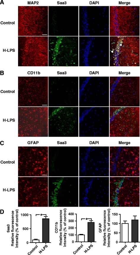
Fig. 2.

Immunofluorescence staining of Saa3 in the CA region of the hippocampus. Frozen sections from the left hemisphere of mice were stained for Saa3 protein using a rabbit anti-Saa3 antibody and Alexa Fluor 488-conjugated anti-rabbit IgG (green fluorescence). The sections were subsequently stained for neurons (anti-MAP2; a), microglia (anti-CD11b; b), or astrocytes (anti-GFAP; c), all in red fluorescence (see the “Methods” section for detail). Cell nuclei were stained with DAPI (blue fluorescence). Immunofluorescence was detected using a confocal laser-scanning microscopy. Images shown are representative of multiple experiments (n = 4 for each group; scale bar, 50 μm). Arrowheads in the merged image mark the positions of the double-stained cells. d quantification of the tissue expression level of Saa3, CD11b, and GFAP after LPS stimulation (15 mg/kg, 24 h). The results are expressed as the means ± SEM from at least three mice per group, each in duplicates or triplicates. *p < 0.05
