Fig. 3.

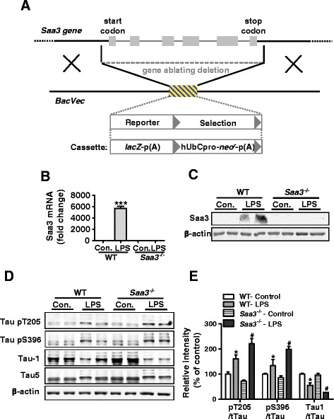
Genetic deletion of Saa3 promotes LPS-induced tau hyperphosphorylation. a The Saa3 gene knockout construct based on information provided by www.komp.org. Gray boxes are exons, and the hatched area denotes expression-selection cassette. b, c Absence of LPS-induced expression of the Saa3 transcript (b) and protein (c), in the Saa3 −/− mice. The brain tissue from the hippocampi of WT and Saa3 −/− mice was collected 24 h after receiving LPS (15 mg/kg) or saline (control). Tissue homogenate was made for real-time PCR (b) and Western blotting (c). Representative blots were probed with an anti-Saa3 antibody. β-actin was used as a loading control.***p < 0.01 compared with mice receiving normal saline (n = 4). d Immunoblots of protein extracts from the hippocampi of WT and Saa3 −/− mice receiving LPS (15 mg/kg, 24 h) or saline (control). Representative blots were probed with anti-phospho-tau antibodies recognizing phosphorylated tau at Thr205 and Ser396 and with antibodies against non-phosphorylated tau (Tau1) and total tau (Tau5), respectively. β-actin was used as a loading control. e Quantification of the immunoblots. The relative phosphorylation level of tau in each sample was normalized against the integrated density of total tau (Tau5) present in each sample. All data shown are mean ± SEM, with four mice in each group; *p < 0.05 compared with WT controls; # p < 0.05 compared with WT receiving LPS stimulation
