Fig. 5.

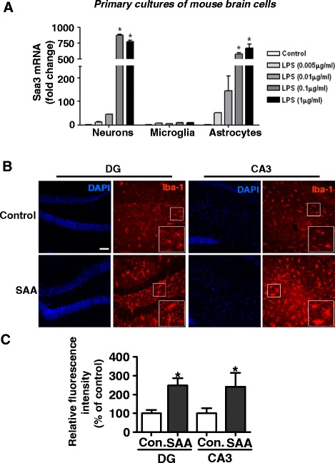
Microglia activation after intracerebral injection of SAA. a LPS-induced Saa3 expression in primary cultures of neurons and astrocytes but not microglia. Quantitative real-time PCR detection of Saa3 transcript using primary cultures of neurons, microglia, and astrocytes after LPS stimulation (5, 10, 100, and 1000 ng/ml) for 24 h. Shown are relative mRNA expression levels based on three independent experiments; mean ± SEM; *p < 0.05 versus controls). b Iba1 stain of the dentate gyrus and CA3 regions of the mouse brain, showing increased microglia activation, based on Iba 1 expression, in mice receiving brain injection of 20 μg SAA compared to those receiving normal saline. c Quantification of Iba1 density, based on images in b and similar samples, was expressed as means ± SEM (three sections per animal; three images at dentate gyrus and CA3 per animal; n = 4 per group; *p < 0.05 compared with the controls (normal saline). Scale bar, 75 μm
