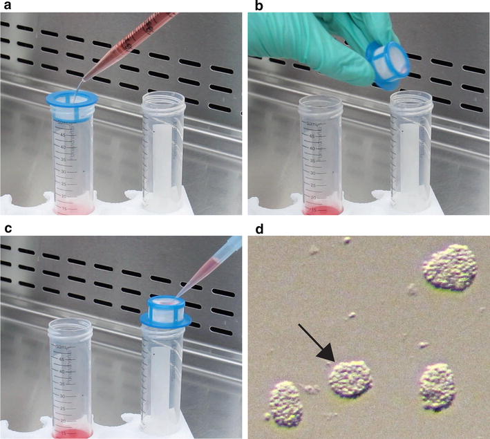
Fig. 1.

Pictorial demonstration on how to obtain OPCAs from cell suspensions. a The cell suspension collected from shaken mixed glial cultures is gently added to a 40 µm cell strainer over a 50 mL conical tube. b The strainer is then flipped upside-down and placed over a new 50 mL conical tube. c Migration media is used to backwash the strainer, causing the transfer of the OPCAs to the conical tube. d An image obtained through the eyepiece of a stereomicroscope, showing freshly isolated OPCAs. When selecting OPCAs for migration experiments, it is desirable to select those most uniform in dimension, such as the one denoted by the black arrow
