Fig. 6.

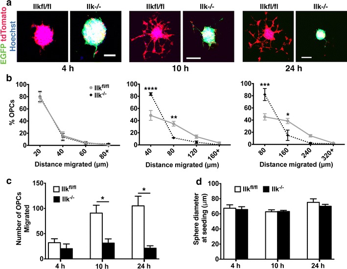
Loss of ILK perturbs OPC migration on fibronectin substrate. a Confocal micrographs of Ilk fl/fl and Ilk −/− OPCAs migrated on Fn for 4, 10 and 24 h. b Quantification revealed no difference in the distance migrated between Ilk −/− and Ilk fl/fl populations at 4 h. However, at 10 h, Ilk −/− OPCs predominantly migrated to the 40 μm proximal ring, and fewer reached the more distant 80 μm ring as compared to Ilk fl/fl cells. Similarly, at 24 h, Ilk −/− OPCs accumulated in the 80 μm proximal ring, with fewer attaining the more distal 160 μm ring. c There was no difference in the total number of migrated Ilk −/− and Ilk fl/fl OPCs at 4 h, while significantly fewer Ilk −/− OPCs had migrated at the 10 and 24 h time points. d The initial diameter of Ilk −/− and Ilk fl/fl OPCAs prior to assay commencement was not significantly different for any of the time intervals investigated. Data represent the mean ± SEM (n = 3). For b, *p < 0.05; **p < 0.01; ***p < 0.001; ****p < 0.0001 (repeated measures two-way ANOVA followed by Bonferroni post-tests). For c and d, *p < 0.05 (Student’s t test). Scale bars, 50 μm for 4 h, 100 μm for 10 and 24 h
