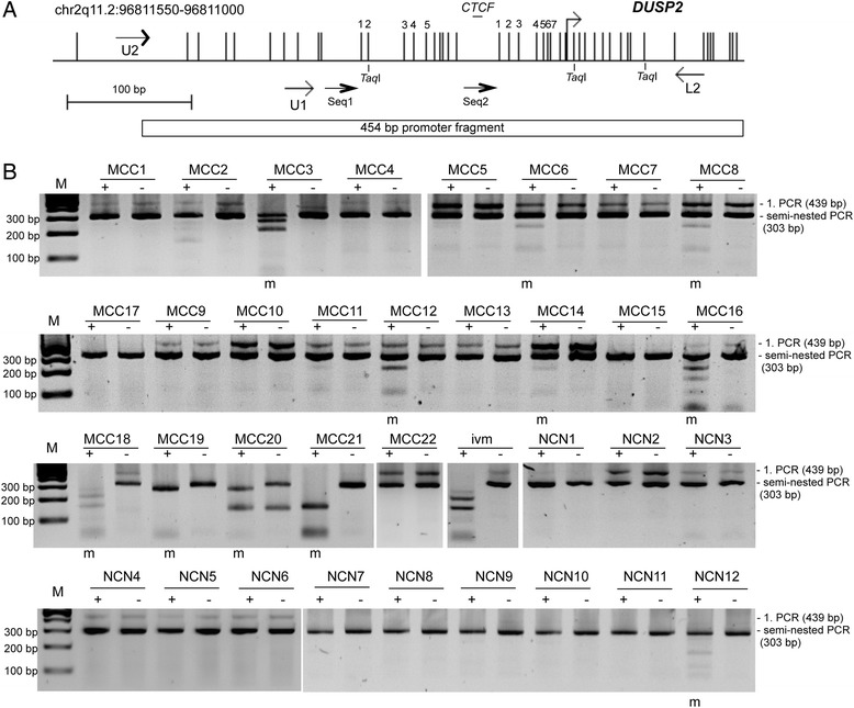
Fig. 1.

Hypermethylation of DUSP2 in primary Merkel cell carcinoma (MCC). a. Structure of the DUSP2 CpG island promoter on chromosome 2q11.2. Vertical lines indicate CpGs and the transcriptional start site is marked. A CTCF motif sequence (GGCAGAGCA; CTCFBSDB2.0) is marked [47]. Primers used for COBRA and sequencing (Seq1 and Seq2) are depicted by arrows. TaqI restriction sites for COBRA and CpGs analyzed by pyrosequencing are indicated. The 454 bp DUSP2 fragment for the luciferase promoter assay is indicated. b. Methylation of DUSP2 in MCC (m = methylated). For COBRA bisulfite-treated DNA from MCC, benign nevus cell nevi (NCN) and in vitro methylated DNA (ivm) was amplified by semi-nested PCR. First and second PCR products are indicated (439 bp and 303 bp, respectively). Products were digested with TaqI (+) or mock digested (-) and resolved on 2 % agarose gels with a 100 bp marker (M)
