Fig. 2.

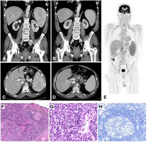
Histology of core biopsy from supraclavicular lymph node and radiological findings including a diagnostic FDG-positron emission tomography (PET) scan and comparative abdominal computed tomography before and after rituximab monotherapy. a PET scan performed during Admission #5 demonstrates widespread avid lymphadenopathy prior to the diagnosis of multi-centric Castleman’s disease (MCD). Images (b) and (c) show splenomegaly on computed tomography (CT) also during Admission #5. Images (d) and (e) were taken following four doses of rituximab therapy. The spleen has decreased in size from a maximum length of 15.5 cm (superioinferiorly) to 12.9 cm. f Low power showing regressed germinal centre and expanded interfollicular zones (H&E, x 100). g High power of interfollicular zone showing plasma cell proliferation (H&E, x 400). h Immunohistochemical staining for HHV-8 shows nuclear expression in isolated cells in the mantle zone (x 200)
