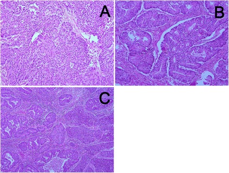
Fig. 2.

Microscopic findings of the tumors. a: The NEC component shows solid sheets and irregular gland like structures with vesicular nuclei and prominent nucleoli. Necrosis and rosette formation are noted. b: The WDEA component shows irregular tubular structures with cribriform pattern and complex folds. c: Histological transition is observed at the boundary between the NEC and WDEA components. Original magnification: a, b: ×200, c: ×100
