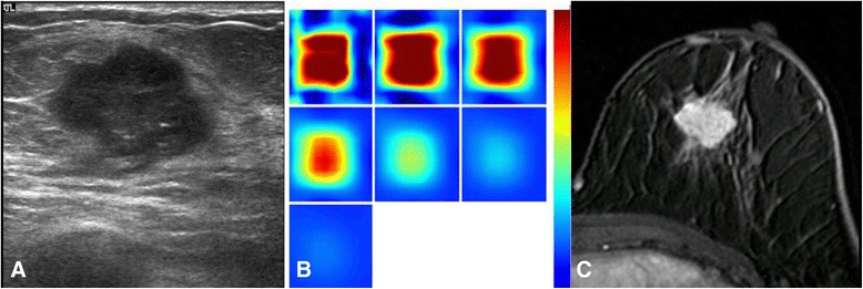
Fig. 1.

A woman with invasive ductal carcinoma (a) A gray-scale ultrasound image shows a hypoechoic mass with microlobulated margins, measuring 1.8 cm in diameter (high histologic grade, LVI (−), ER(−), PR(−), HER-2 (+), Ki-67 (+)). b A reconstructed optical absorption map shows a distinct mass with a high maximum THC of 293.4 μmol/L. The first section (slice 1, top left) is a 6 × 6 cm spatial x-y image (coronal plane of the body) obtained at a depth of 0.5 cm, as measured from the skin surface. The last section (slice 7, bottom left) is a 6 × 6 cm spatial x-y image (coronal plane of the body) obtained at a depth of 3.5 cm, as measured from the skin surface. Spacing between sections is 0.5 cm in the direction of propagation. c A lobular homogenously enhancing mass is noted from one of the DCE-MRI slices. The K trans is 0.122 [1/min], the k ep is 0.415 [1/min] and the SER is 1.024
