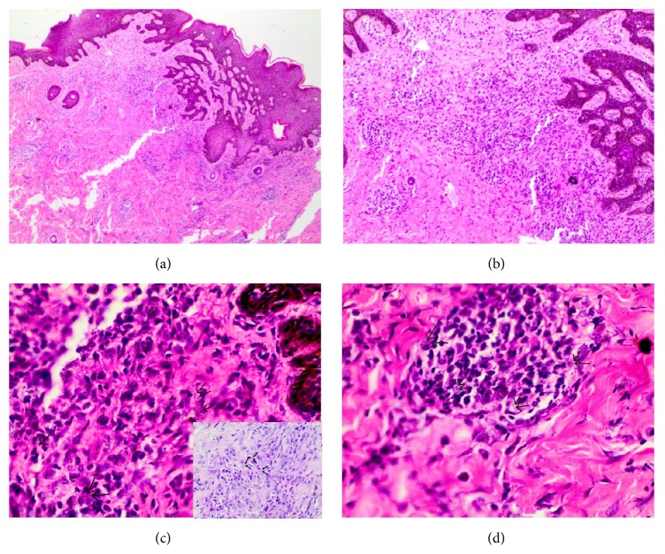
Figure 2.
(a) Diffuse inflammatory infiltrate composed of chronic inflammatory cells and macrophages (H&E, ×40); (b) yeast-like forms widely dispersed in the dermis (H&E, ×100); (c) yeast-like forms widely dispersed in the dermis, black arrows pointing out the spores (H&E, ×400). Insert shows a period acid-Schiff stain with arrows pointing out the fungal spores; (d) a granuloma composed of macrophages containing S. schenckii organisms (H&E, ×400).

