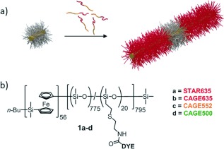
Figure 1.

Fluorescent cylindrical micelles of PFS56‐b‐PDMS775/DYE20. a) Schematic representation of the formation of fluorescent cylindrical micelles by seeded growth from a non‐fluorescent seed (not to scale); yellow=PFS core, red=fluorescent corona, grey=non‐fluorescent corona. b) Chemical structure of PFS56‐b‐PDMS775/DYE20, showing the position of the fluorescent dyes (DYE).
