Fig. 1.

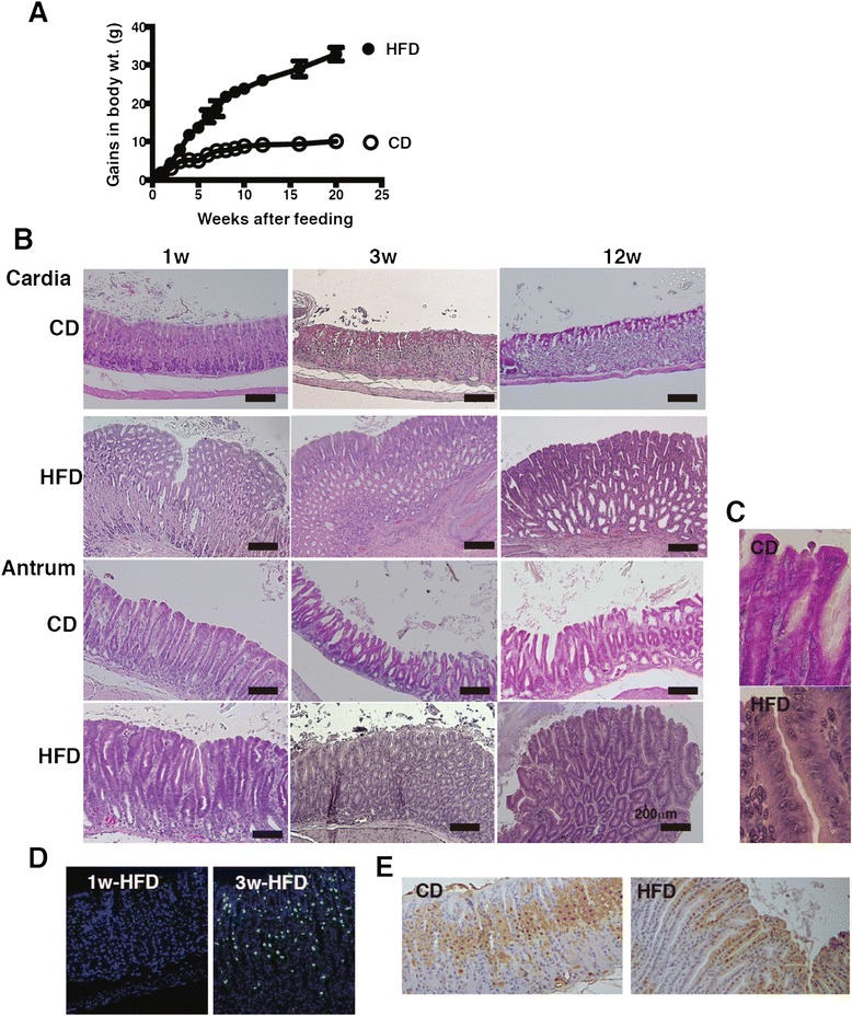
Pathological changes of gastric mucosa owing to HFD-feeding. a Alteration of gains in body weights of C57BL/6 J mice fed CD (n = 10) or HFD (n = 10) during 20 weeks. b Representative H&E-sections of the gastric cardia and antrum from mice fed CD or HFD for 1, 3, and 12 weeks. c Magnified image of the gastric antrum in mice fed CD and HFD in Fig. 1b at 12 weeks after feeding (magnification, ×400). The cell nucleolus, nuclear hypertrophy, dyspolarity, and pseudostratification were observed. d CD45 staining of the gastric mucosa of 1 and 3 week HFD-fed mice. e Ki67-staining in the gastric mucosa of mice fed CD or HFD for 3 weeks. 5–10 mice were used in each analysis, and representative data are shown
