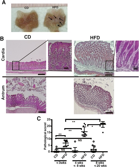
Fig. 2.

Development of gastric mucosal atrophy in diet-induced obese mice. a The gastric lumen was opened along the outer curvature of mice fed CD or HFD for 20 weeks. Arrows indicate the polyp-like lesions in the stomach of HFD-fed mice. b Representative H&E-sections of the gastric cardia and antrum from mice fed CD or HFD for 20 weeks. c The histological scores from the stomachs of mice fed CD or HFD (<3 weeks, 4–8 weeks, 8–20 weeks of feeding; 10 mice per group) were graded according to the diagnostic criteria described in the Methods. Results were analyzed by the Kruskal-Wallis test, followed by a Dunn’s multiple comparison test. * p < 0.05, ** p < 0.01, NS; not significant
