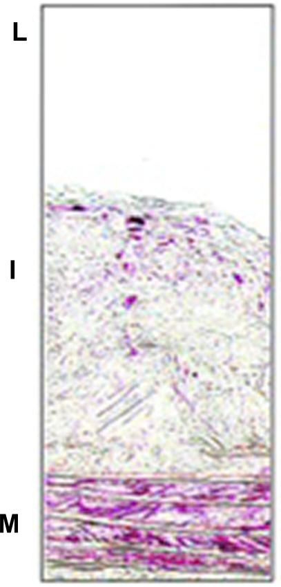
Figure 3.
Immunostaining of smooth muscle α-actin ( red stain) in atherosclerotic lesions of ApoE −/− mice fed western diet for 16 weeks. L indicates lumen; I, intima; M, media. Note the two positive areas: at the bottom of the section, where the bands of the internal elastic laminae are visible, are the VSMC in the medial layer (M) of the artery. In the intimal area (I) are VSMC that migrated from the media and continue to express smooth muscle α-actin. From Rong JX et al. Circulation.2001; 104:2447-2452.

