
Figure 3.
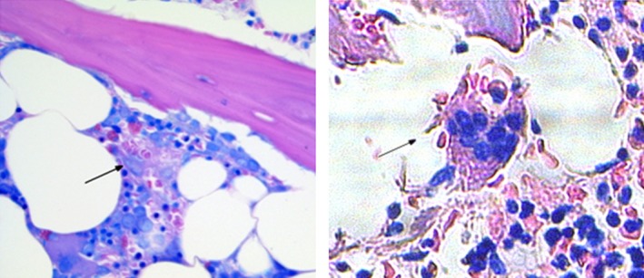
Giemsa‐stained bone marrow specimens obtained at diagnosis of hemophagocytic lymphohistiocytosis (HLH) (left) and at autopsy about 2 months later (right). Images show macrophages with erythro‐ (left and right) and leukophagocytic (right) activity representing key features of HLH disease activity.