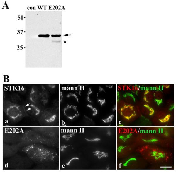
Figure 1. Wild-type STK16 distributes to the Golgi while E202A is present on peripheral puncta.
(A) Cell lysates from control (uninfected), STK16- or E202A-expressing cells were immunoblotted with anti-V5 antibodies. Molecular mass markers are indicated on the left-hand side in kDa. The arrow is pointing to the 35 kDa wild-type and E202A STK16 species. The band marked with an asterisk is probably a partially degraded E202A. (B) WIF-B cells expressing STK16 (a–c) or E202A (d–f) were double-labelled for mannosidase II (mann II) and the V5 tag. Merged images are shown in c and f. Scale bar, 10 μm.

