
Fig. 1.
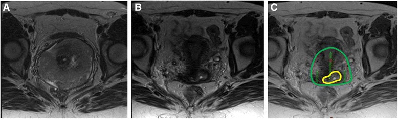
Axial T2 weighted MR images. a: Pre-treatment image with large cervical tumor. b: Pre-brachytherapy image showing excellent response to treatment with small focus of residual signal intensity. c: Simulated brachytherapy plan with GTV contoured (yellow) and plan adjusted to cover residual disease with 100 % isodose line (green) using tandem and ovoid applicators