
Fig. 6.
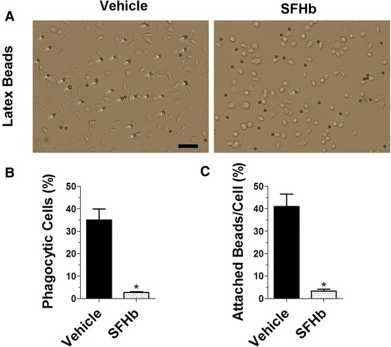
Hemoglobin treatment inhibits microglial phagocytosis. Primary microglial cells were treated with SFHb (1 mM) or vehicle for 2 h and then incubated with fluorescent latex beads (diameter 6 μm) for 1 h. Representative images were taken and white arrows illustrate the attached beads to microglial cells (a). Bar graphs show that the hemoglobin pretreatment markedly reduces the percentage of phagocytic microglial cells (b) and the numbers of attached beads (c). Scale bar, 50 μm. Values represent means ± SEM. Differences between two groups were determined by unpaired two-tailed Student’s t test. The experiment was repeated three times and in n ≥ 150 cells