Fig. 1.

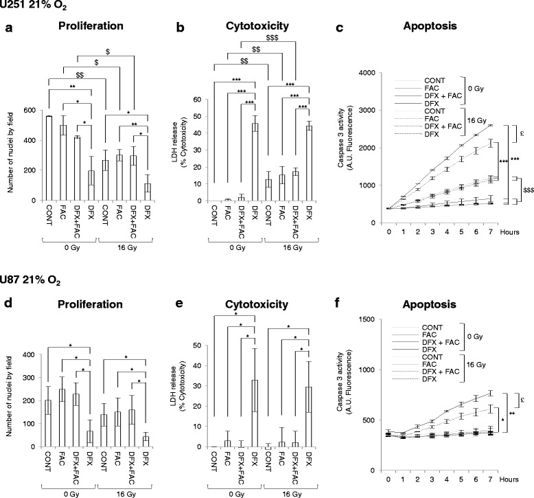
Deferasirox inhibits proliferation linked with increased cytotoxicity and apoptosis in glioblastoma cells under normoxic conditions. Number of nuclei of U251 (a) and U87 (d) glioblastoma cells cultivated at 21 % of oxygen in non-treated condition (CONT) or 3 days after treatment with 5 μM of ferric ammonium citrate (FAC), or with 10 μM of deferasirox and 5 μM of FAC (DFX + FAC) or with 10 μM of deferasirox (DFX) in non-irradiated condition (0 Gy) or following irradiation with 16 Gy (16 Gy). The number of nuclei are expressed as mean ± standard deviation (S.D.) (n = 3). Measure of lactate dehydrogenase (LDH) release into cell culture medium of U251 (b) and U87 (e) glioblastoma cells cultivated at 21 % of oxygen in untreated condition (CONT) or 3 days after treatment with 5 μM of ferric ammonium citrate (FAC), or with 10 μM of deferasirox and 5 μM of FAC (DFX + FAC) or with 10 μM of deferasirox (DFX) in non-irradiated condition (0 Gy) or following irradiation with 16 Gy (16 Gy). Cytotoxicity is expressed as mean percentage ± standard deviation (S.D.) (n = 3) of the total amount of LDH released from cells and relative to glioblastoma cells treated with 0.1 % Triton X-100, given the arbitrary percentage of 100. DEVD-AMC caspase 3 activity in U251 (c) and U87 (f) glioblastoma cells cultivated at 21 % of oxygen in untreated condition (CONT) or 3 days after treatment with 5 μM of ferric ammonium citrate (FAC), or with 10 μM of deferasirox and 5 μM of FAC (DFX + FAC) or with 10 μM of deferasirox (DFX) in non-irradiated condition (0 Gy) or following irradiation with 16 Gy (16 Gy). Caspase 3 activity is expressed as mean arbitrary units (A.U.) of fluorescence per 30 μg of proteins ± standard error of the mean (SEM) (n = 3). One-way ANOVA was performed between DFX treatment and CONT, FAC or DFX + FAC conditions in non-irradiated (0 Gy) or irradiated (16 Gy) conditions (*, p-value ≤0.05; **, p-value ≤0.01; ***, p-value ≤0.001). Two-way ANOVA was performed between non-irradiated (0 Gy) condition and irradiated (16 Gy) condition ($, p-value ≤0.05; $$, p-value ≤0.01; $$$, p-value ≤0.001). Two-way ANOVA was performed between in DFX treatment in non-irradiated condition (0 Gy) and irradiated (16 Gy) condition (£, p-value ≤0.05)
