


Figure 1. Spontaneous liver injury and hepatic steatosis is present in 14 weeks old Arg2−/− mice.
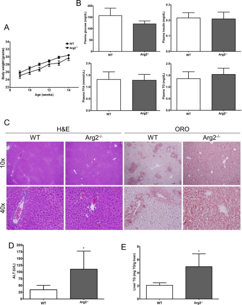
Body weight (A) along with several metabolic parameters – plasma glucose level, plasma insulin level, levels of free fatty acids in plasma, plasma triglycerides – are comparable in Arg2−/− mice and WT mice fed with normal chow (B). However, analysis of liver histology and assessment of hepatic steatosis with Oil Red O staining uncovered the development of liver injury and hepatic steatosis (C) (10× and 40×). In line with this ALT, levels were significantly increased in Arg2−/− mice when compared to WT mice (D). The amount of liver triglycerides was also increased in Arg2−/− (Arg2−/− 4.9 vs. WT 2.6 mg TG/g liver, p <0.05, E). Mean ± S.E.M. are shown. *p<0.05.