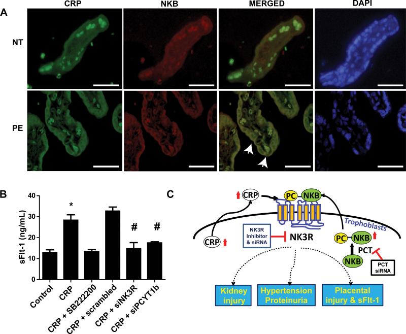
Figure 5. CRP and NKB are localized in the syncytiotrophoblast cells of placentas of PE patients and CRP directly induces sFlt-1 secretion from cultured term human villous explants via NK3R signaling in a PCT-dependent manner.
(A) Coimmunofluorescence revealed increased CRP and NKB in syncytiotrophoblast cells in terminal villi. On the merged image, membrane colocalization was visualized along the syncytiotrophoblast cells. Some autofluorescence can be seen intravascularly due to the presence of autofluorescent RBCs. (40x magnification; scale bar = 100 μm). (B) Placental villus explants isolated from normal patients and treated with or without CRP in the presence or absence of series of drugs or siRNA. CRP directly induced secretion of sFlt-1 from cultured human villus explants, but this increase was attenuated by treatment with SB222200 or siRNA for NK3R or PCYT1b. * = p < 0.05 difference from control; # = p < 0.05 difference from CRP + scrambled. (n = 4 wells placental villous explant culture) (C)Working model: CRP is expressed in the placentas and elevated CRP in the syncytiotrophoblast cell is additional source underlying increased circulating CRP in PE patients. Locally-synthesized CRP cross-talks with post-translationally modified phosphocholinated NKB (PC-NKB) by placental-specific enzyme, PCT. Subsequently, CRP and PC-NKB work together preferentially activates NK3R leading to increased sFlt-1 secretion. Our findings reveal novel mechanisms for pathogenesis of PE and identify innovative therapeutic possibility for prevention and treatments.

