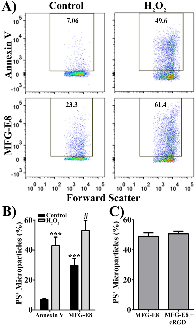
Figure 3.
Hydrogen peroxide increases presence of phosphatidylserine on the MP surface. (A) Representative scatter plots of MPs from control and 500 μM H2O2-treated RPE cells. Microparticles were stained with either annexin V or MFG-E8 to measure phosphatidylserine-positive (PS+) MPs. (B) Quantification of PS+ MPs measured by two PS-binding proteins, annexin V and MFG-E8. (C) Quantification of MFG-E8 binding to MPs from H2O2-treated RPE cells in the absence (MFG-E8) of or in the presence (MFG-E8 + cRGD) of cyclic RGD. Data are presented as mean ± SD (n = 3). ***P < 0.0001 compared with control annxein V; #P < 0.002 compared with control MFG-E8.

