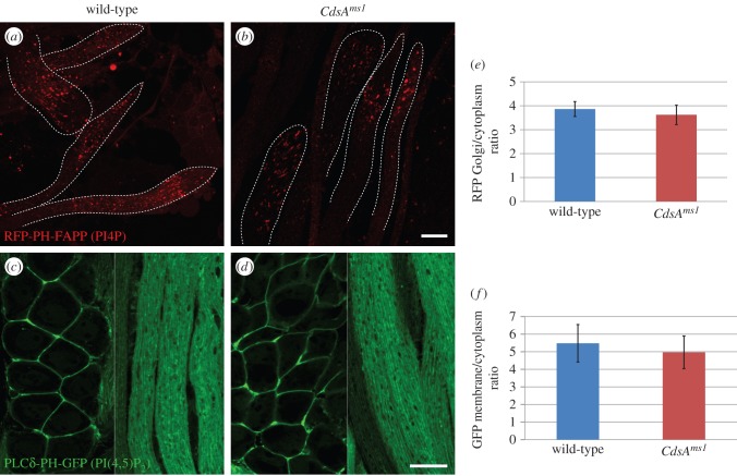
Figure 5.
Distribution of phosphoinositides in CdsAms1 male germ cells. (a,b) Confocal micrographs of the squashed and fixed preparations of elongated spermatids expressing the PI4P-binding RFP-PH-FAPP transgene. RFP-PH-FAPP localizes to punctate structures both in the wild-type (a) and CdsAms1 (b) elongated spermatids. (c–f) Confocal micrographs of male germ cells expressing PLCδ-PH-GFP, which binds PI(4,5)P2. In spermatocytes (left side of images) and in elongated spermatids (right side of images), PI(4,5)P2 is localized to the plasma membrane in wild-type (c) and CdsAms1 (d) testes. Images were taken with the same exposure time in the case of each transgene, with or without the CdsAms1 homozygotes. Membrane to cytoplasmic RFP intensity (e) or GFP intensity (f) ratios were calculated from measurements of mean pixel intensities within the same sized area of membrane versus cytoplasm. Scale bars, 20 µm.

