Figure 7.
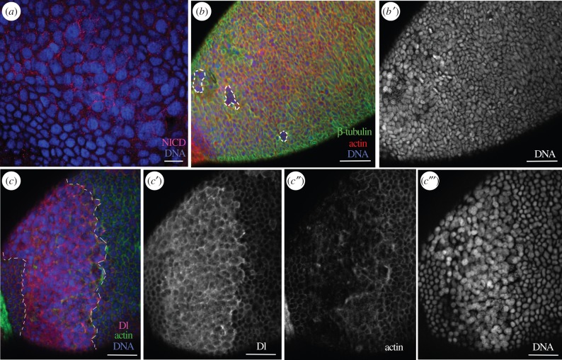
The BgN receptor and BgDl ligand determine the differentiation of posterior FCs. In 5-day-old dsBgHpo-treated adult females, the FCs in the posterior pole showed changes in their morphology and overexpressed NICD (a). Some FCs in this area lacked β-tubulin and F-actin (b, dashed frames). (b′) The nuclei of the latter cells were round in shape and bigger. (c–c‴) BgDl overexpression in the band of FCs (between the dashed lines), sourrounding the posterior FCs (c); (c′) Dl labelling was detected in the posterior FCs where usually it is not present; (c″) actin staining; shows the microfilaments clearly affected in the posterior region and (c‴) the nuclei of these FCs were bigger and with a differential staining intensity for DAPI compared to the rest of FCs. The F-actin microfilaments were stained with phalloidin-TRITC (red in b and green in c), DNA with DAPI (blue). The posterior pole of the basal follicle is orientated towards the bottom left. Scale bars, 20 µm in (a) and 50 µm in (b,c).

