
Figure 2.
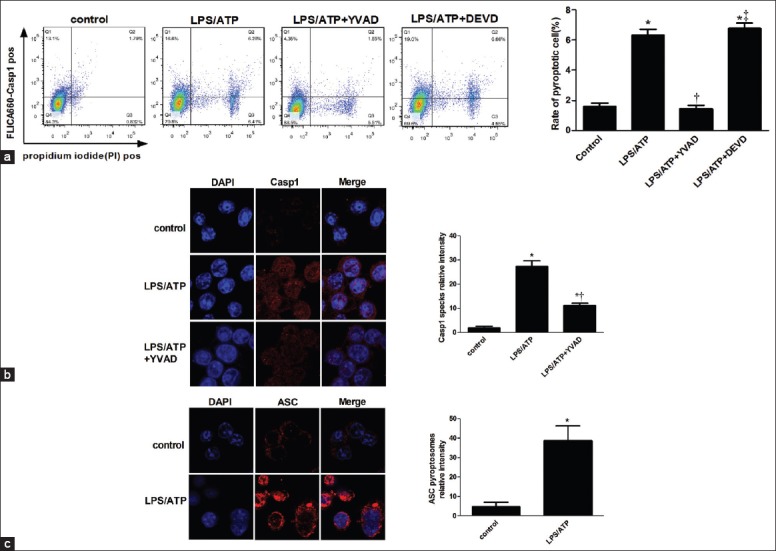
Alveolar macrophage pyroptosis occurs ex vivo after LPS stimulation. Alveolar macrophages isolated from mice were stimulated for 5 h with or without LPS (500 ng/ml) and ATP (5 mmol) added during the last hour of culture, in the absence or presence of 50 μmol of YVAD or DEVD. (a) Cells were stained with FLICA, and pyroptotic cells were detected by flow cytometry. Caspase-1 speck (b) and ASC (c) cells were analyzed by immunofluorescence. *P < 0.05 vs. the control group; †P < 0.05 vs. the LPS/ATP group. ‡P < 0.05 vs. the lipopolysaccharide/ATP + YVAD group. Results are representative of three separate independent experiments.